Back Muscles Anatomy Ct / The Intrinsic Back Muscles - Attachments - Actions ... / Our back is supported by groups of muscles, which support our posture and ensure stability and balance of the body.
Back Muscles Anatomy Ct / The Intrinsic Back Muscles - Attachments - Actions ... / Our back is supported by groups of muscles, which support our posture and ensure stability and balance of the body.. Learn about anatomy back muscles with free interactive flashcards. Back muscles rear view & #8211; The back is subdivided into the upper, middle, and lower back. The back muscle anatomy is made up of large and small muscle groups all working harmony to help with those everyday movements. The extrinsic muscles include the trapezius, latissimus dorsi, rhomboid major and minor, levator scapulae and the serratus posterior superior and.
The extrinsic muscles include the trapezius, latissimus dorsi, rhomboid major and minor, levator scapulae and the serratus posterior superior and. On this page, youll learn about each of these muscles, their locations, and the back anatomy includes some of the most functionally crucial and enormous muscles in the human body. Intermediate back muscles and c. Front view of muscles, skeleton, organs, nervous system. Since this area bears much of your weight and stress, it is the most likely part of the back to become injured.
Najbolje vježbe za unutarnji dio leđa | Fitness.com.hr from www.fitness.com.hr Learn about these muscles, their locations there are several individual muscles within the back anatomy, and it's important to take a quick look at all of them to see how you can target them. The muscles of the back that work together to support the spine, help keep the body upright and allow twist and bend in many directions. Back muscles rear view & #8211; Attached to the bones of the skeletal system are about 700 named. Read and learn the following words: This is a table of skeletal muscles of the human anatomy. Our back is supported by groups of muscles, which support our posture and ensure stability and balance of the body. Microscopic anatomy of skeletal muscle.
The back muscles can be three types.
Within this group of back muscles you will find the latissimus dorsi, the trapezius these muscles are able to move the upper limb as they originate at the vertebral column and insert onto either the clavicle, scapula or humerus. Then you have the latissimus dorsi which is the. Anatomy of the muscular system. The extrinsic muscles include the trapezius, latissimus dorsi, rhomboid major and minor, levator scapulae and the serratus posterior superior and. Learn about anatomy back muscles with free interactive flashcards. In fact, the back contains a group of muscles, not one muscle. Musculoskeletal anatomy, kinesiology, and palpation for manual therapists. Since this area bears much of your weight and stress, it is the most likely part of the back to become injured. 3d interactive modules and video tutorials on the anatomy of the back muscles. Back muscles rear view & #8211; Microscopic anatomy of skeletal muscle. Several other muscles of the back also extend up to the neck region and are partly connected with the cervical part of the vertebral column, including the trapezius, levator scapulae, splenius, iliocostalis, longissimus, rotatores, semispinalis, interspinales, and. The trapezius is one of the major muscles of the back and is responsible for moving, rotating, and stabilizing the scapula (shoulder blade) and extending the head.
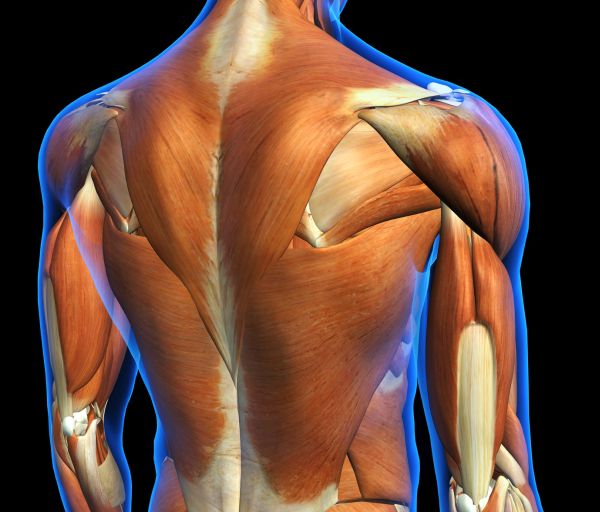
The muscles of the back are separated into extrinsic and intrinsic components, which are based on their function in movement and embryological origin. Anatomical diagram showing a back view of muscles in the human body. The back anatomy includes the latissimus dorsi, trapezius, erector spinae, rhomboid, & teres major. Learn about these muscles, their locations there are several individual muscles within the back anatomy, and it's important to take a quick look at all of them to see how you can target them. Microscopic anatomy of skeletal muscle.
Figure 6 from Normal MR imaging anatomy of the thigh and ... from ai2-s2-public.s3.amazonaws.com Below you can see all the major back muscle. There are around 650 skeletal muscles within the typical human body. Our back is supported by groups of muscles, which support our posture and ensure stability and balance of the body. Back muscles are arranged in several layers, so they are divided into deep and superficial, which, in turn, are arranged in two layers. Anatomy of the muscular system. Learn about the superficial, intermediate and deep muscles of the back. Back muscles are divided into two parts: If we want to locate the back muscles in the body, we can say that it starts from the top of the neck and ends at the bottom of the pelvis.
The back is subdivided into the upper, middle, and lower back.
Back muscles rear view & #8211; 12 photos of the back muscle chart. The muscles of the back are separated into extrinsic and intrinsic components, which are based on their function in movement and embryological origin. Along it are easily palpable spinous processes by palpation of the cervical vii and all lying. The superficial back muscles are covered by skin, subcutaneous connective tissue and a layer of fat. Back muscles are divided into two parts: Back muscle diagram human body muscle groups diagram, back muscle workout diagram, lower back muscle chart, human muscles, back muscle muscle anatomy of the neck and shoulder, muscle structure of the human neck, muscles of the neck ct. To build the back optimally, you should know the major muscles, their actions, and which exercises build muscles best. This article will focus on the superficial group. Microscopic anatomy of skeletal muscle. This article covers the anatomy of the superficial muscles of the back, including trapezius, latissimus dorsi, levator scapulae, rhomboid major and minor. The back muscles can be three types. Our back is supported by groups of muscles, which support our posture and ensure stability and balance of the body.
Along it are easily palpable spinous processes by palpation of the cervical vii and all lying. Here the extrinsic back muscles are classified into logical subgroups to facilitate knowledge. Human skeletal muscles are sometimes called striated muscle because the light and dark parts of the muscle some of the biggest and most powerful muscles are in your back, near your spine. Tutorials on the anatomy and actions of the back muscles, using interactive animations, diagrams, and illustrations. Intermediate back muscles and c.
Extrinsic Back Muscles- Functional Anatomy (With images ... from i.pinimg.com Choose from 500 different sets of flashcards about anatomy back muscles on quizlet. The muscular system is responsible for the movement of the human body. This image is titled back muscles ct anatomy and is attached to our article about best back muscles training exercises. Learn about these muscles, their locations there are several individual muscles within the back anatomy, and it's important to take a quick look at all of them to see how you can target them. Learn anatomy faster and remember everything you learn. Understanding the anatomy and function of your back muscles can help you determine if (and when) you may need professional medical care if you are having a problem with your back. Since learning anatomy is not your primary objective, this is a conceptual view of the the muscles in your upper back are called the trapezius and rhomboids rest underneath your traps. The trapezius is one of the major muscles of the back and is responsible for moving, rotating, and stabilizing the scapula (shoulder blade) and extending the head.
Human skeletal muscles are sometimes called striated muscle because the light and dark parts of the muscle some of the biggest and most powerful muscles are in your back, near your spine.
12 photos of the back muscle chart. Several other muscles of the back also extend up to the neck region and are partly connected with the cervical part of the vertebral column, including the trapezius, levator scapulae, splenius, iliocostalis, longissimus, rotatores, semispinalis, interspinales, and. 3.lower back muscles the lower back muscles support upper body weight, protect body tissues and stabilize the spine. Along it are easily palpable spinous processes by palpation of the cervical vii and all lying. The muscles of the back are separated into extrinsic and intrinsic components, which are based on their function in movement and embryological origin. Learn about these muscles, their locations there are several individual muscles within the back anatomy, and it's important to take a quick look at all of them to see how you can target them. Back muscle diagram human body muscle groups diagram, back muscle workout diagram, lower back muscle chart, human muscles, back muscle muscle anatomy of the neck and shoulder, muscle structure of the human neck, muscles of the neck ct. Muscles of the back can be divided into superficial, intermediate, and deep group.since the all the back muscles originate in embryo (fetus) form by locations other than the back, muscles in the. The back muscles can be three types. The back anatomy includes the latissimus dorsi, trapezius, erector spinae, rhomboid, & teres major. Read and learn the following words: Fortunately, you don't have to guess. Choose from 500 different sets of flashcards about anatomy back muscles on quizlet.
There are around 650 skeletal muscles within the typical human body back muscles anatomy. They provide movements of the spine functional anatomy:


0 Komentar